Saturday, March 21, 2009
Friday, March 20, 2009
Exercise tracing2
1

2

3

4

5

6

7

8

9 10
10
 10
10
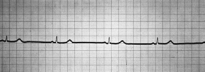
rhythm strip 2---------Ventricular Asystole
rhythm strip 3---------1st degree heart block
rhythm strip 4---------SVT
rhythm strip 5---------2nd degree heart block mobitz type II
rhythm strip 6---------3rd degree herat block
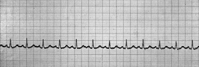
rhythm strip 7---------Atrial Flutter
rhythm strip 8--------Atrial Fibrillation
rhythm strip 9--------sinus tachycardia
rhythm strip 10-------VT
Friday, March 6, 2009
Exercise tracing 4
Wednesday, March 4, 2009
Basic ECG rhthym
Sinus rhythm
 sinus rhythm has the following Criteria:
Rate should range between 60-100, regular, there is P wave preceding each complex ,the PR interval is 3-5 small square ,and QRS complex is narrow 1-3 small square
sinus bradycardia
 sinus bradycardia Everything is normal except that rate less than 60/min.
 Everything is normal except that rate more than 100 up to 150/min.
 The above rhythm is VT has same criteria like SVT with exception of the width of the complex it is wide bizarre shape (regular rhythm rate 150-250 no P wave and complex is wide)
VF
 Disorganized chaotic electrical signals this VF This patient needs to be defibrillated!! QUICKLY
Asystole
 Looking at the ECG you'll see that:
Rhythm - Flat
Rate - 0 Beats per minute
QRS Duration - None
P Wave - None so this asystole carry on CPR
 fixed block One beat will be conducted other not conducted so some P wave followed by QRS complex other P wave not followed by P wave
 No relation between P wave and QRS complex (p wave regular at normal rate and QRS complex is regular at slower rate but no relation between them
Junctional Rhythms

 Regular rhythm with inverted P wave or no P wave note that the complex is narrow at rate of 40-60 /min.
IDIOVENTRICULAR rhythm
 sinus rhythm has the following Criteria:
Rate should range between 60-100, regular, there is P wave preceding each complex ,the PR interval is 3-5 small square ,and QRS complex is narrow 1-3 small square
sinus bradycardia
 sinus bradycardia Everything is normal except that rate less than 60/min.
sinus tachycardia
 Everything is normal except that rate more than 100 up to 150/min.
PAC (premature atrial CONTRACTION) 
VT

Occasional irregularity with some beats came early in position followed by pause look like the preceding wave narrow in nature (beat number 3 &6)
PVC (premature ventricular CONTRACTION)
 Occasional irregularity with some beats came early in position followed by pause but wide bizarre shape so the difference between PAC and PVC is is the width of the complex
 the above rhythm is AF it is totally irregular and no P wave
 Occasional irregularity with some beats came early in position followed by pause but wide bizarre shape so the difference between PAC and PVC is is the width of the complex
AF
 the above rhythm is AF it is totally irregular and no P wave
 The above rhythm is VT has same criteria like SVT with exception of the width of the complex it is wide bizarre shape (regular rhythm rate 150-250 no P wave and complex is wide)
VF
 Disorganized chaotic electrical signals this VF This patient needs to be defibrillated!! QUICKLY
Asystole
 Looking at the ECG you'll see that:
Rhythm - Flat
Rate - 0 Beats per minute
QRS Duration - None
P Wave - None so this asystole carry on CPR
now we talk about heart blocks we have three types of heart block first ,second and third but the second degree has two subtypes type one and two first degree heart block
 the above tracing is first degree heart block fixed prolonged PR interval above 5 small square
second degree type 1
 2nd Degree Block Type 1 (Wenckebach or Mobitz type I)progressive prolongation of PR interval until P had no QRS complex
second degree type 2
 fixed block One beat will be conducted other not conducted so some P wave followed by QRS complex other P wave not followed by P wave
Third degree heart block
 No relation between P wave and QRS complex (p wave regular at normal rate and QRS complex is regular at slower rate but no relation between them
Junctional Rhythms

 Regular rhythm with inverted P wave or no P wave note that the complex is narrow at rate of 40-60 /min.
IDIOVENTRICULAR rhythm

Rate: 40 bpm
Rhythm: Regular
P wave: no look
the complex is wide
paced rhythm

Artificial spike before each complex
Subscribe to:
Comments (Atom)


 VT
VT


 Atrial Flutter


 6.
6.

 1. Paced rhythm
2. Second degree type 1
3. Atrial flutter
4. Asystole
5. VF
6. AF
7. First degree block
 Most of the time it is regular rhythm with flutter wave like saw tooth appearance

